
Mechanism-led bioelectric repair
The corneal epithelium maintains a transepithelial potential through active ion transport and barrier integrity. When the surface is wounded, local barrier interruption converts that potential into wound-directed ion current and electric fields. Corneal epithelial cells can respond to physiologic electric fields with directional migration.
Human donor-cornea studies show the corneal wound current is measurable and can be increased pharmacologically. GALVANIS uses this biology as a screening system: move the pharmacodynamic signal first, then test whether closure improves in the same model.
How we decide
A pharmacodynamic readout of the wound's repair current, paired with functional closure across models.
Advancement rule
Signal first, spend second.
Endogenous electric fields (EFs)
Wound currents create lateral fields oriented into the defect.

Quantifiable pharmacodynamics (PD)
Measure ΔTEP / Δion flux and relate to closure kinetics.

GO/NO GO Gates
Advance only candidates meeting PD + closure thresholds.


Bioelectricity in corneal wound repair
The corneal epithelium maintains a transepithelial potential difference (TEP/TEPD) via active ion transport and tight junction resistance. When the epithelium is wounded, resistance drops and the TEP partially collapses (“short-circuit”), driving ionic currents toward the defect. Those currents generate lateral electric fields oriented into the wound, and corneal epithelial cells migrate directionally in physiologic electric fields.
Selected references: Reid et al., Cornea (2011); Vieira / Reid et al. (2011); Zhao et al., Nature (2006); Zhao et al., Cold Spring Harbor Perspectives in Biology (2022).
OcuRegen™: a topical bioelectric modulator built for corneal repair
Galvanis is building PD-gated topical bioelectric therapies for corneal repair. Corneal wounds generate measurable endogenous electric currents, and human donor corneas show those currents can be chemically modulated. Our pre-seed program does not bet the company on a single modality belief. Only candidates that preserve barrier function, increase corneal wound TEP, and improve closure advance. That gives OcuRegen™ a faster developmental path.
BIOELECTRIC MECHANISM
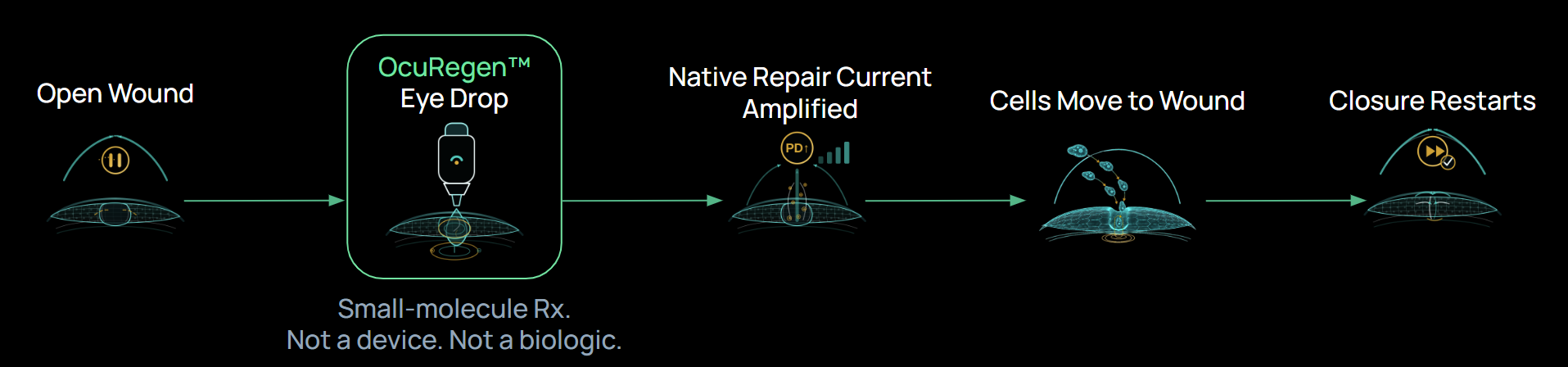
Designed to amplify the wound's endogenous repair current and restore its electric field.

PHARMACODYNAMIC GATED DE-RISKING
Advance only candidates meeting pre-specified PD thresholds (ΔTEP) plus functional closure improvement ex vivo.

ADDITIVE WITH CURRENT CARE
Designed to be used alongside existing therapies to further accelerate healing.

OPHTHALMOLOGY-READY
Preservative-free formulation goals with standard ocular safety/tolerability gating.

Restore the wound’s electric field to guide closure.
Corneal wounds generate endogenous electric fields that drive directional epithelial migration. In impaired healing, the electric field is reduced, slowing closure. OcuRegen™ is designed to amplify the wound's endogenous repair current and accelerate directional epithelial migration toward the injury.
Pharmacodynamic readout of the wound's repair current.
Lead selection gated by that signal plus faster closure across models.
Goal: a clinically meaningful acceleration of closure versus vehicle and an active comparator (preclinical benchmark).
A name with lineage: Luigi Galvani
In the late 1700s, Italian physician Luigi Galvani observed that a frog’s leg could twitch in response to electrical stimulation; an early window into what he called “animal electricity.” His work helped launch electrophysiology and shaped how scientists think about bioelectric signals in living tissue. GALVANIS is a nod to that lineage: living tissues generate electric signals, and in modern biology we can measure and engineer those signals.